Figure 2.

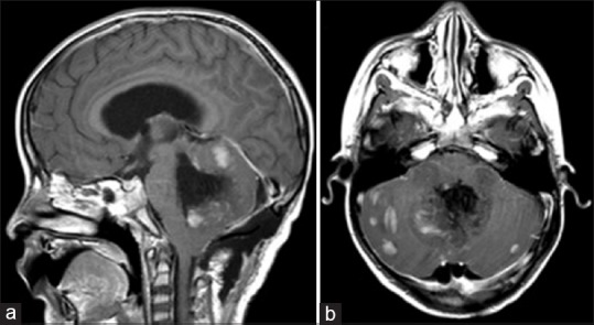
Postoperative MRI Brain, T1 post-gadolinium in sagittal (a) and axial (b) sections. The medial portion of the lesion has been largely resected, but the lesion remains in the bilateral cerebellar hemispheres, totaling >1.5 cm2

Postoperative MRI Brain, T1 post-gadolinium in sagittal (a) and axial (b) sections. The medial portion of the lesion has been largely resected, but the lesion remains in the bilateral cerebellar hemispheres, totaling >1.5 cm2