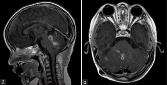
Figure 3.

Post-radiotherapy MRI Brain, T1 post-gadolinium in sagittal (a) and axial (b) sections. There has been some interval improvement of leptomeningeal spread and nodular lesions. However, there has been recurrence of disease in the fourth ventricle
