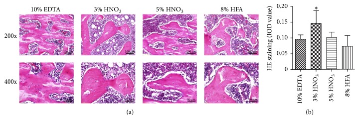
Figure 2.
Representative images (a) and histological images analysis (b) of HE staining showed the influences of different decalcified solutions on staining quality of rat femurs. Image-Pro Plus was used to quantify the relative IOD value of HE staining of the trabecular bone. ∗p < 0.05 compared with EDTA group.

