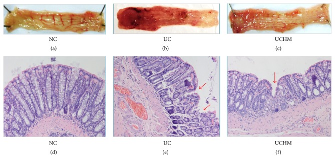
Figure 4.
Colon tissue injuries in all groups. (a) and (d) were both NC groups. (b) and (e) were both UC groups. (c) and (f) were both UCHM groups. (a), (b), (c) represent the changes of the representative colon observed by the naked eye. (d), (e), (f) show representative HE staining (HE, ×200) of the histopathological changes of colon tissues under light microscopy.

