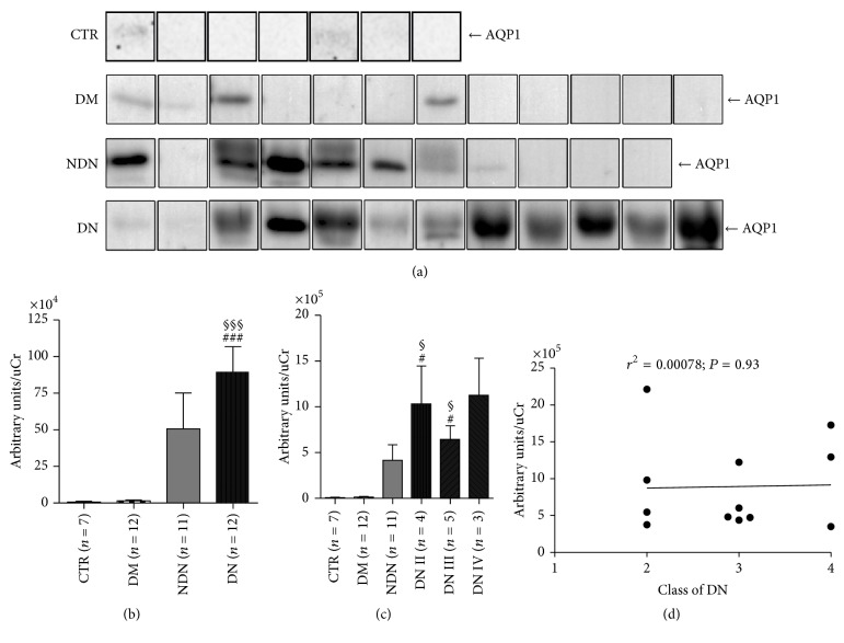
Figure 4.
The urinary excretion of AQP1 increases in both DN and NDN patients and does not correlate with the clinical severity of DN. (a) Exosomes were isolated from urine of healthy subjects (CTR, n = 7), patients with DM (n = 12), patients with NDN (n = 11), and patients with DN (n = 12) using the two-step differential centrifugation method. Total exosome proteins were resolved on 12% SDS-PAGE and analyzed by Western blotting for AQP1 abundance. (b) Densitometry analysis of the uAQP1 band intensities was normalized for uCr and reported as means ± SEM. §§§P < 0.001 versus CTR and ###P < 0.001 versus DM, obtained by Kruskal-Wallis one-way ANOVA test. (c) DN patients were grouped according to the histological changes evaluated by kidney biopsy: DN stage II (n = 4), DN stage III (n = 5), and DN stage IV (n = 3) and densitometry analysis of uAQP1 at each stage compared to uAQP1 in CTR and DM and NDN patients. All data are reported as mean ± SEM. §P < 0.05 versus CTR and #P < 0.05 versus DM, obtained by Kruskal-Wallis one-way ANOVA test. (d) Linear regression analysis of uAQP1 abundance, as semiquantified by Western blotting, with the class of DN (r2 = 0.00078; P = 0.93).

