
Figure 5. Effects of taxol on axon regeneration in rats after nerve injury surgery.
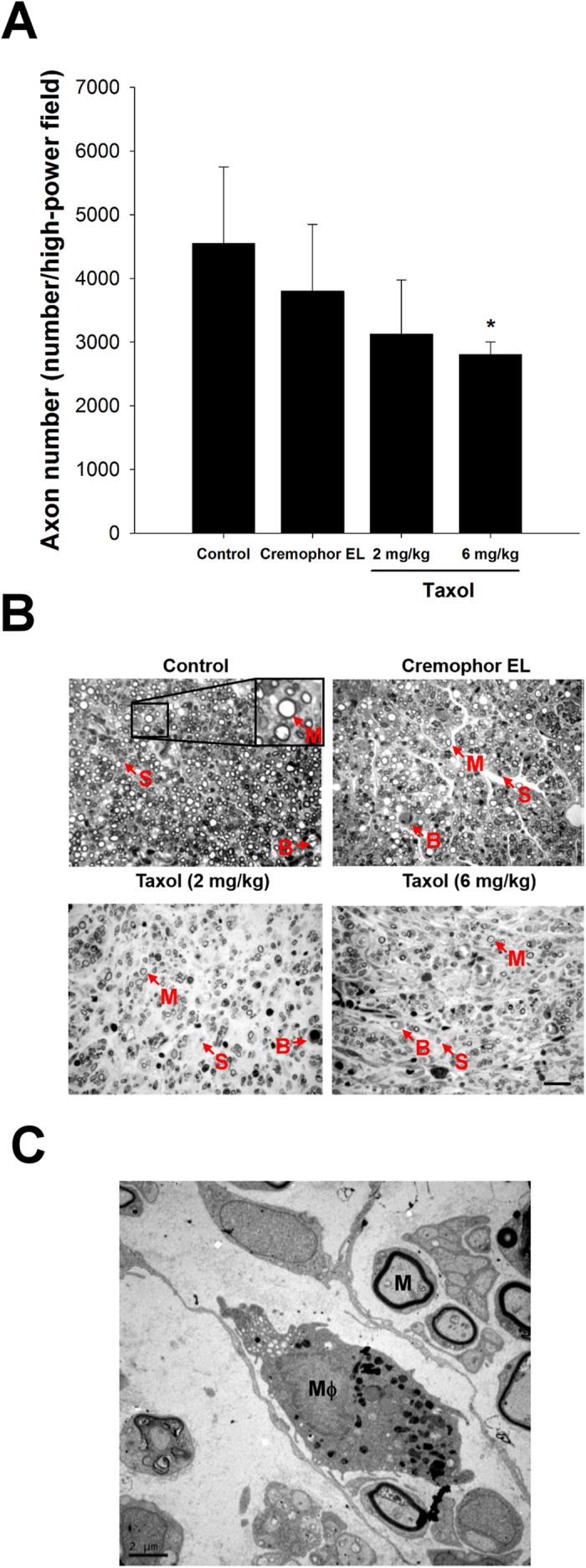
(A) Quantitation of myelinated axonal counts in regenerated sciatic nerve cross-sections. (B) Representative histological micrographs of nerve tissue (M: myelinated axon; S: Schwann cell; B: blood vessel). (C) Ultrastructural analysis using electron microscopy to determine the macrophage (Mϕ) infiltration and remyelination in taxol-treated groups. The values represent means ± standard deviation (SD) for 10 rats for each group. *P < 0.05, compared to control and Cremophor EL groups. Scale bars = 20 μm and 2 μm for (B) and (C), respectively.