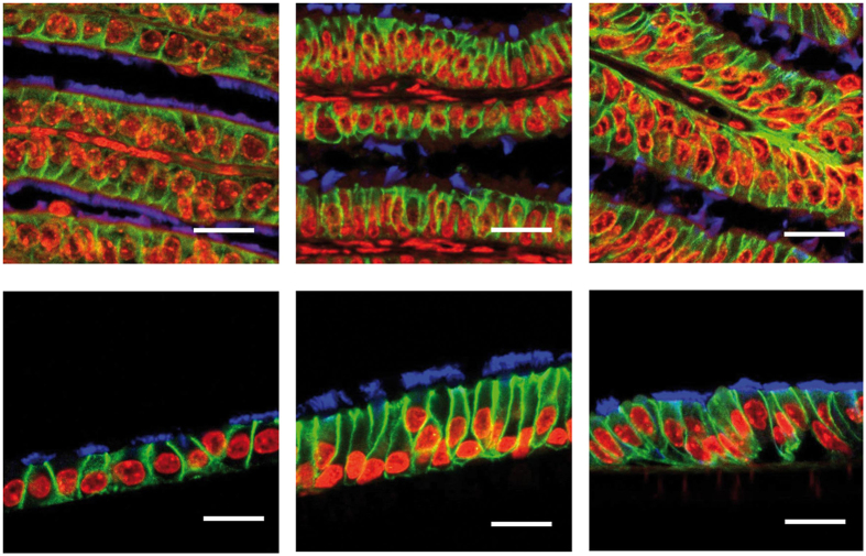
Figure 2. Oviduct epithelial cells of different mammalian species grown at the air-liquid interphase (ALI).
Immunodetection of epithelial markers in murine, porcine and bovine oviduct tissue (from left to right; upper pictures) and respective ALI-OEC (lower pictures) after 21 d (ALI-MOEC and -POEC) or 28 d (ALI-BOEC) of culture. Red: nuclei; green: beta-catenin (cell-cell adhesions); blue: acetylated tubulin (cilia); bar = 10 μm.

