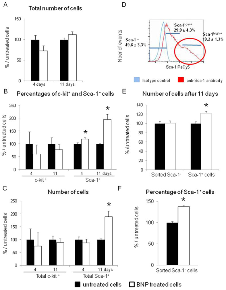
Figure 1. BNP stimulates Sca-1+ cell proliferation.
(A) Non myocyte cells (NMCs) were isolated from neonatal hearts of C57BL/6 mice, cultured 4 and 11 days with or without BNP (untreated cells) and counted. (B) Percentages of c-kit+ and Sca-1+ cells obtained by flow cytometry analysis on BNP treated or untreated NMCs. (C) Number of cells expressing the c-kit or the Sca-1 protein in NMCs treated or not with BNP for 4 and 11 days calculated with the total number of cells and the percentages of the c-kit+ and Sca-1+ cells. (A–C) n = 8 and 16 different experiments after 4 and 11 days of culture, respectively. (D) Representative histogram of NMC sorting for Sca-1 expression. The numbers represent the percentage of the cells compared to the total number of sorted NMCs. n = 18–43 different experiments. (E) Number of sorted Sca-1− and Sca-1high+ cells treated or not with BNP for 11 days. n = 6 and 12 for Sca-1− and Sca-1high+ cells, respectively. (F) Percentages of Sca-1+ cells among sorted Sca-1− cells treated or not with BNP for 9 days. n = 4 different experiments. (A–E) All results expressed as fold-increase above the results obtained in untreated cells. All the results are means ± SEM. *p < 0.05.

