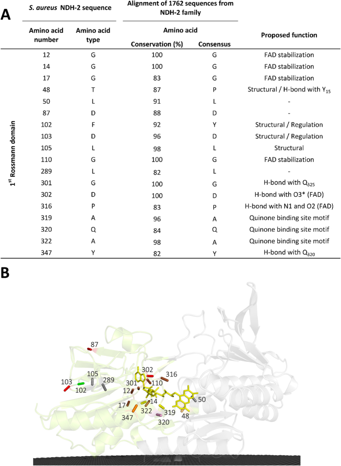
Figure 2. Amino acid residue conservation in the first dinucleotide binding domain.
(A) List of the 18 amino acid residues present in the 1st dinucleotide (FAD) binding domain that are conserved in at least 80% of the NDH-2s; (B) Cartoon representation of the X-ray crystal structure of NDH-2 from S. aureus (PDB:4XDB7) highlighting the location of the 18 conserved amino acid residues present in this domain. Membrane is represented in black.

