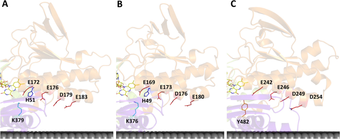
Figure 6. Proton pathways in the second dinucleotide binding domain.
Cartoon representations of the X-ray crystal structures highlighting the proton pathways proposed for NDH-2s from (A) S. aureus (PDB:4XDB7); (B) C. thermarum (PDB:4NWZ6); (C) S. cerevisiae (PDB:4G734). Membrane is represented in black.

