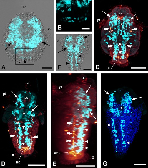
Figure 1.

EdU and immunocytochemical labeling of solenogaster developmental stages. Maximum intensity projections of confocal image stacks; apical/anterior is up in all panels; scale bar equals 50 μm in (A) and (C–G) and 20 μm in (B). (A) Labeling of nuclei of proliferating cells (light blue) with overlay of transmitted light image; early larva of Gymnomenia pellucida showing kidney‐shaped apical proliferation zones (arrows) and anlage of longitudinal bands of proliferating cells (arrowhead). (B) Detail of boxed area in (A) without overlay of transmitted light image. (C) Labeling of nuclei of proliferating cells (light blue) and serotonin‐like immunoreactive (LIR) components of the nervous system (orange); 12–13 days posthatching (dph) larva of Wirenia argentea scanned in ventral aspect showing anterior proliferation zones (arrows) with gap in the region of the foregut (asterisk), longitudinal bands of proliferating cells (arrowheads), and longitudinal (lateral) neurite bundles (double arrowheads). (D) Labeling of nuclei of proliferating cells (light blue) and serotonin‐LIR components of the nervous system (orange); 16–17 dph larva of W. argentea scanned in ventral aspect showing anterior proliferation zones (arrows) with gap in the region of the foregut (asterisk), longitudinal bands of proliferating cells (arrowheads), and longitudinal (lateral) neurite bundles (double arrowheads). (E) Labeling of nuclei of proliferating cells (light blue) and serotonin‐LIR components of the nervous system (orange); 3D reconstruction (volume rendering, MIP (max) mode) of the same confocal image stack as in (D) in right lateral view. Note that the lateral neurite bundles (double arrowheads) are located dorsally to the longitudinal bands of proliferating cells (arrowheads). Serotonin‐LIR elements at the same level as the longitudinal bands of proliferating cells belong to the developing ventral nervous system. The asterisk marks the region of the foregut and arrows point to the anterior proliferation zones. (F) Labeling of nuclei of proliferating cells (light blue) with overlay of transmitted light image; late larva of G. pellucida scanned in ventral aspect showing anterior proliferation zones (arrows) with gap in the region of the foregut (asterisk) and longitudinal bands of proliferating cells (arrowheads). (G) Labeling of nuclei of proliferating cells (light blue) and cell nuclei with DAPI (dark blue); late larva of G. pellucida scanned in left ventrolateral aspect showing anterior proliferation zones (arrows) with gap in the region of the foregut (asterisk) and longitudinal bands of proliferating cells (arrowheads). ao, apical organ; at, apical tuft; pt, prototroch; src, suprarectal commissure; tt, telotroch.
