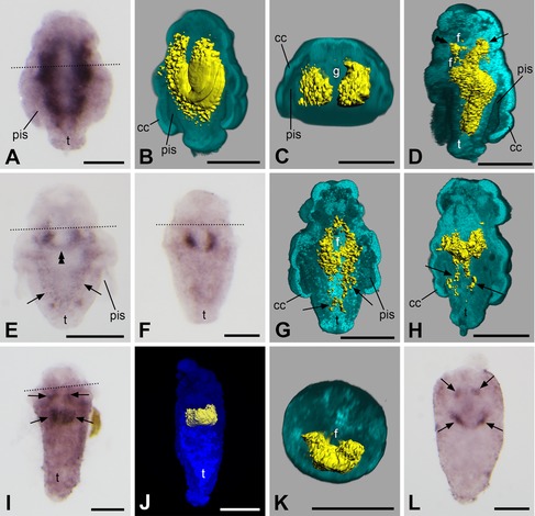
Figure 4.

Expression of twist in larvae and juveniles of Wirenia argentea. Apical/anterior is up in (A), (B), (D–J), and (L); dorsal is up in (C) and (K); scale bar equals 50 μm in all panels; dotted lines indicate the region of the prototroch. (A) 10–11 days posthatching (dph) larva. (B) 3D reconstruction of a confocal scan with volume rendering (blend mode) of autofluorescence of larva (light blue) and surface rendering of reflection signal of expression domain (yellow); 6–11 dph larva in left ventrolateral view with ventral part of autofluorescence signal omitted. Note that the gene expression signal does not extend to the outermost cell layer of the larva. (C) Same specimen and reconstruction as in (B) in apical/anterior view with apical/anterior part of autofluorescence signal omitted. Note that the gene expression signal is restricted to the subepidermal parts of the trunk and that it is absent in both the calymma and the gut. (D) 3D reconstruction of a confocal scan with volume rendering (blend mode) of autofluorescence of larva (light blue) and surface rendering of reflection signal of expression domain (yellow); 10–11 dph larva in left lateral view with left part of autofluorescence signal omitted. Note the bifurcation of the anterior part of the expression domain with one branch encompassing the foregut (double arrowhead) and another branch extending in anteriodorsal direction (arrow). The gene expression signal does not extend to the outermost cell layer of the larva. (E) 14–15 dph larva. Note the ventral fusion of the expression domain (double arrowhead) and the remnants of the lateral bands in the trunk (arrows). (F) 12–13 dph larva. (G) 3D reconstruction of a confocal scan with volume rendering (blend mode) of autofluorescence of larva (light blue) and surface rendering of reflection signal of expression domain (yellow); 14–15 dph larva in left dorsolateral view with dorsal part of autofluorescence signal omitted. Note that the expression domain encircles the foregut. Remnants of its lateral bands in the trunk are still visible (arrows) and expression is purely subepithelial. (H) 3D reconstruction of a confocal scan with volume rendering (blend mode) of autofluorescence of larva (light blue) and surface rendering of reflection signal of expression domain (yellow); 10–11 dph larva in left ventrolateral view with ventral part of autofluorescence signal omitted. Note that remnants of the lateral bands of the expression domain are still visible in the trunk (arrows) and that twist expression is purely subepithelial. (I) 14–15 dph larva showing twist expression domain (arrows) encircling the foregut. (J) 3D reconstruction of a confocal scan with volume rendering (MIP (max) mode) of autofluorescence of larva (dark blue) and surface rendering of reflection signal of expression domain (yellow); 14–15 dph larva in ventral view. (K) 3D reconstruction of a confocal scan with volume rendering (blend mode) of autofluorescence of larva (light blue) and surface rendering of reflection signal of expression domain (yellow); same specimen as in (J) in apical/anterior view with apical/anterior part of autofluorescence signal omitted. Note that twist expression is purely subepithelial. (L) 16–18 dph juvenile showing twist expression domain (arrows) encircling the foregut. pis, peri‐imaginal space; cc, calymma cell; f, foregut; g, gut; t, outgrowing trunk.
