
Figure 3.
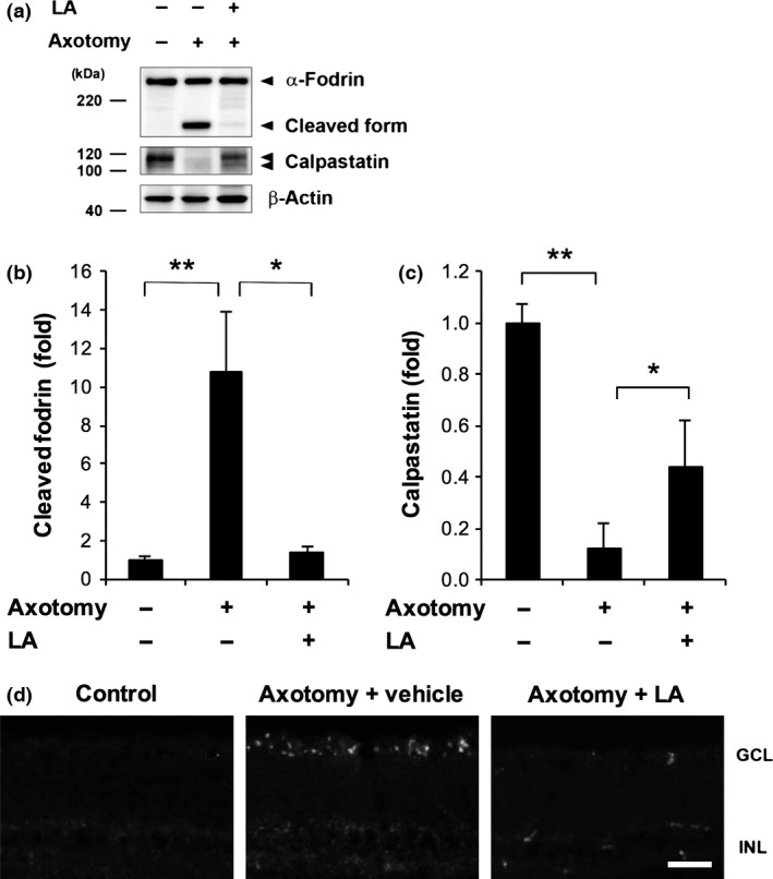
Latanoprost acid (LA) inhibited calpain activation and calpastatin breakdown after optic nerve transection. (a) LA (200 pmol/eye) inhibited the axotomy‐induced degradation of α‐fodrin, a prominent substrate for μ‐calpain, and calpastatin. The full‐length and cleaved forms of α‐fodrin are estimated to comprise 240 and 150 kDa peptides, respectively. Beta‐actin was used as an internal control. (b and c) Quantitative analysis of axotomy‐induced calpain activation and calpastatin breakdown in whole retinas 7 days after axotomy. The signal intensity of each band was normalized to 1 in the non‐treated group. (*p < 0.05, **p < 0.01, n = 5 in each group). Error bars denote SD. (d) Representative fluorescence images of frozen sections showing the results of treatment with a fluorogenic cell‐penetrating calpain substrate (2 μL of 100‐μM) in the eyes. With LA treatment, the signal of the cleaved calpain substrate, which was mainly detected in the GCL, was lower on day 7 after axonal injury. The vehicle was saline containing 5% dimethylsulfoxide. GCL, ganglion cell layer; INL, inner nuclear layer; ONL, outer nuclear layer. Scale bar = 50 μm.