
Figure 4.
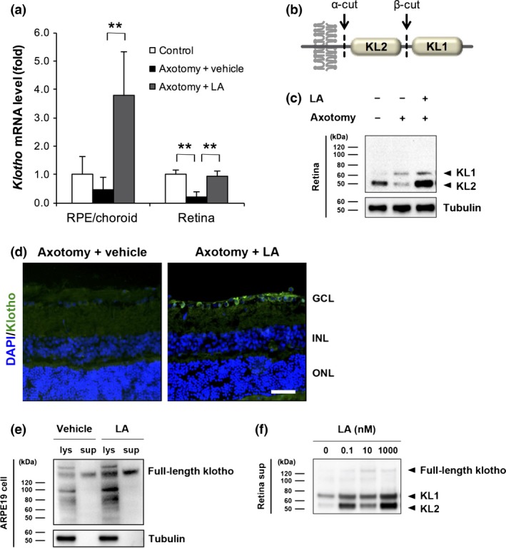
Latanoprost acid (LA)‐mediated promotion of klotho expression and shedding in the retinal pigment epithelium (RPE) and retina. (a) Three days after optic nerve transection, the intravitreal injection of LA (200 pmol/eye) significantly increased the mRNA level of klotho in the RPE/choroid and retina. The vehicle was saline containing 5% dimethylsulfoxide (DMSO). Gapdh was used as an internal control (**p < 0.01, n = 4 in each group). Error bars denote SD. (b) Schematic diagram of the transmembrane klotho structure and klotho shedding. The extracellular domain of klotho protein is composed of two homologous domains, KL1 and KL2. The extracellular domain of klotho possesses two cleavage sites (α‐cut and β‐cut), with three types of fragments (KL1, KL2 and soluble full‐length klotho) being produced extracellularly as a result of klotho shedding. (c) LA inhibited the post‐injury reduction in KL2 in the retina and increased the protein level of klotho on day 3. The level of KL1 in the axotomized rats did not significantly differ with or without LA. Tubulin was used as an internal control. (d) Immunostaining of klotho in retinal sections 3 days after optic nerve transection in LA‐treated rats. GCL, ganglion cell layer; INL, inner nuclear layer; ONL, outer nuclear layer. Scale bar = 50 μm. (e) In human ARPE19 cells, the application of LA at 1 nM for 24 h promoted the expression and shedding of full‐length klotho in both cell lysate (lys) and culture supernatant (sup). The vehicle was a culture medium containing 0.1% DMSO. Tubulin was used as an internal control. Neither KL1 nor KL2 were observed in the ARPE19 cells. (f) The promotion of klotho shedding from primary retinal cells at various concentrations of LA (0.1, 10, and 1000 nM).