
Figure 2.
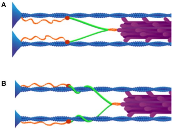
Winding filament hypothesis. (A) Upon Ca2+ influx, N2A titin (red) binds to thin filaments (blue). (B) Cross-bridges (purple) wind PEVK titin (green) on thin filaments in active muscles. As shown, all titins in the same half-sarcomere must wind in the same direction around actin filaments. Reproduced with permission from Nishikawa et al. (2012).