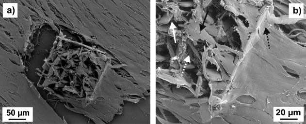
Figure 8.

SEM images at two different magnifications of a cage microstructure with 35 µm square pores on a PETG substrate seeded with NBDS cells and cultured in ODM. Both images show the same sample. In (b), the straight white arrow points to part of a pore sidewall, the straight black arrow to a cell inside the structure, the dashed white arrow to an area which is rich in fibrous extracellular material, and the dashed black arrow to a cell part at the outer rim of the structure.
