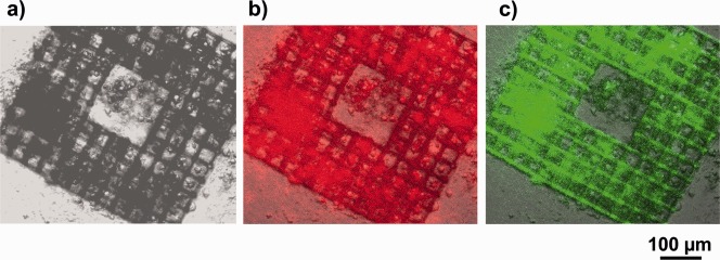
Figure 10.

(a) Bright‐field microscope image of a cage microstructure with 35 µm square pores on a PETG substrate seeded with NBDS cells cultured in ODM; (b) image (a) overlaid with an immunofluorescence image of red fluorescent collagen type I antibodies (same sample); (c) image (a) overlaid with a fluorescence image of FITC‐labeled phalloidin (same sample).
