
Figure 4.
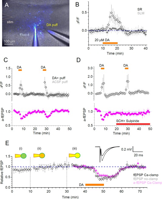
Putative roles of astrocytes in hippocampal circuit dopamine signalling. A: Experimental arrangement in s. lacunose moleculare (SLM), showing recording whole‐cell pipette (Fluo‐4), dopamine pressure application pipette (DA puff), and stimulating electrode (stim) shown; patched astroglia and GJCs stained via gap‐junction escape of Fluo‐4 can be seen. B: Distinct dopamine (DA) sensitivity of astroglia in s. radiatum (SR) and SLM: Time‐course (mean ± SEM) of Ca2+ response to the bath application of 20 μM DA in SR (hollow) and SLM (filled) GJCs, n = 12 and n = 10, respectively; difference is at least at P < 0.05 (unpaired t‐test) over the 15‐20 min window. C: Open circles, time course (mean ± SEM) of Ca2+ ‐dependent (Fluo‐4) fluorescence of GJCs in SLM during two brief (3 min) local DA puffs (200 µM in the puff pipette, as in A), as shown (n = 10); grey filled circles, control experiments with no DA in the puff pipette (n = 7); magenta, time course of a‐fEPSPs recorded from the patched astroglia (n = 3) indicating reversible dopaminergic inhibition of perforant path—CA1 synapses, reported earlier. D: Experiments as in C but with DA receptor blockers 5 µM SCH23390 and 20 µM Sulpiride applied after the first stimulus (red) abolishing synaptic inhibition (bottom, n = 3) but not astroglial Ca2+ responses (top, n = 6); notations as in C. E: Dopaminergic inhibition of perforant path fEPSPs or a‐fEPSPs persists after astrocytic Ca2+ clamp. Open circles, time course (mean ± SEM) of normalized fEPSP slope for cell‐attached (i), whole‐cell equilibrating (ii) and whole‐cell recording (iii) followed by bath application of DA (black bar) (n = 4); magenta, a‐fEPSPs recorded form the patched cell in Ca‐clamp conditions (n = 4); grey filled circles, control fEPSPs, with no Ca‐clamp (n = 6); traces, examples of fEPSPs of varied amplitudes recorded in one experiment. [Color figure can be viewed at wileyonlinelibrary.com]