Figure 3.
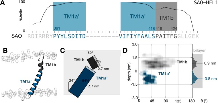
The SAO deletion mutant peptide also samples an ensemble of conformations. (A) The peptide remains mostly helical during the ensemble of 50 filtered simulations. As expected, proline and glycine residues initiate and terminate regions of high helicity. Two putative transmembrane segments were defined, each starting from one of the two proline residues, a central segment (TM1a′) and a shorter segment identical to that found in the wild-type sequence (TM1b). (B) Illustrative snapshot taken from the end of one of the molecular dynamics simulations. The peptide is colored according to panel A. The positions of the phosphate groups of the lipid bilayer are shown as empty circles. (C) To-scale schematic representation of the average conformation with segment tilts, lengths, and helicity annotated. The average extent of the lipid bilayer as defined by the phosphate atoms is shaded gray. (D) Density plot showing the variation in the depth and tilt angle of both segments. The center of mass of the larger transmembrane segment (TM1a′, colored dark blue) remains on average 0.8 nm below the center of the lipid bilayer, and the segment explores tilt angles between 10° and 60°, centered around ∼35°.

