Figure 5.
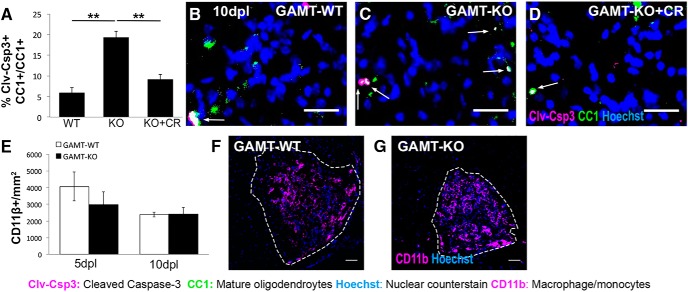
Gamt-deficient oligodendrocytes exhibit reduced survival after focal spinal cord demyelination. A, Quantification of immunostaining showing the proportion of oligodendrocytes positive for cleaved caspase-3 (Clv-Csp3+CC1+) of total oligodendrocytes (CC1+) at 10 dpl in Gamt+/+ (GAMT-WT), Gamt−/− (GAMT-KO), and Gamt−/− mice treated with 25 ng of creatine (GAMT-KO + CR). n = 3 mice/condition; one-way ANOVA with Bonferroni post hoc test. B–D, Representative immunostainings of dying oligodendrocytes in (GAMT-WT) (B), GAMT-KO (C), and GAMT-KO + CR (D) lesions at 10 dpl. White arrows indicate cells double positive for Clv-Csp3 and CC1. Scale bars, 25 μm. E, Quantification of immunostaining for CD11b+ cells per square millimeter in GAMT-WT and GAMT-KO lesions at 5 and 10 dpl. n = 3 mice/condition. F, G, Representative immunostainings of macrophages/microglia (CD11b+; magenta) in GAMT-WT (F) and GAMT-KO (G) lesions at 5 dpl. Scale bars, 50 μm. Data are represented as mean ± SEM. Brightness and contrast were adjusted for visualization. *p < 0.05, **p < 0.01.

