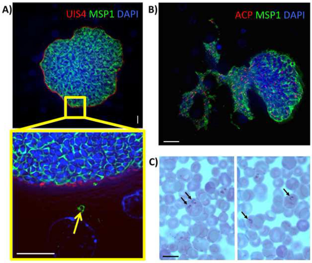
Figure 2. Complete maturation of P. vivax liver stages and persistence of hypnozoites in the FRG KO huHep mouse.
(A) Indirect immunofluorescence assay of P. vivax liver stages at ten days after sporozoite infection using Pv merozoite surface protein-1 (MSP-1) polyclonal rabbit antibody (green), PvUIS4 mouse monoclonal antibody (red) and DAPI (blue, DNA). MSP-1 expression reveals the presence of differentiated exo-erythrocytic merozoites within the liver stage schizont. The magnification of the area in the yellow box shows a MSP-1- positive exo-erythrocytic merozoite (arrow) outside the confines of the mature liver schizont. Scale bar: 10 µm. (B) IFA of a nine-day old P. vivax liver stage stained with PvMSP-1 polyclonal rabbit antibody (green) to visualize the merozoite surfaces, ACP monoclonal mouse antibody (red) to visualize the apicoplast and DAPI (DNA stain, blue). The mature liver stage is in the process of releasing exo-erythrocytic merozoites into the surrounding liver tissue. Scale bar: 20 µm. (C) Human red blood cells enriched for reticulocytes were injected into FRG KO huHep mice nine days post sporozoite infection. Four hours later, the blood was removed and microscopically analyzed by Giemsa-stained thin blood smear. Black arrows point to P. vivax ring stage parasites within reticulocytes. Scale bar: 10 µm.

