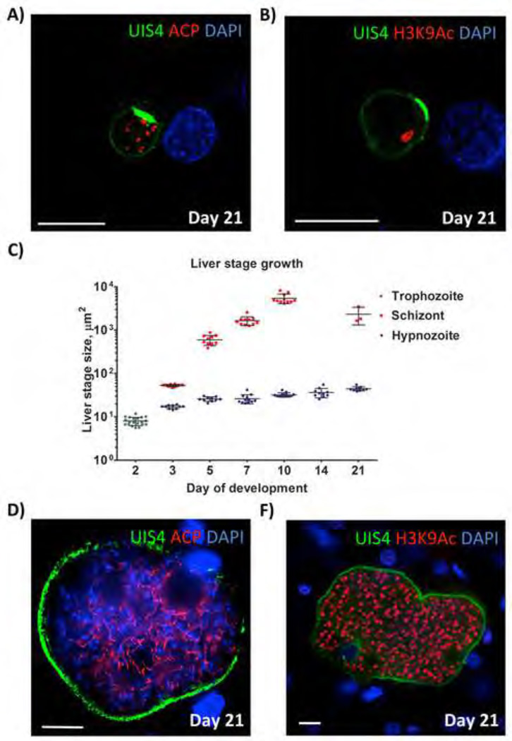
Figure 5. P. vivax hypnozoite persistence and hypnozoite activation in FRG KO huHep mice.
(A, B) Persistent hypnozoites at day 21 after sporozoite infection. Parasites were visualized with antibodies to PvUIS4 (green), antibodies to acetylated lysine 9 of histone H3 (H3K9Ac) (red) and host hepatocyte nuclei were visualized with DAPI. The UIS4-postive PVM prominence is maintained and notable in the PVM of all persistent hypnozoites. Hypnozoites contained a single H3K9Ac-positive structure. (C) Size comparison of liver stage trophozoites, liver stage schizonts and hypnozoites at different time points of infection. The size (liver stage area at the greatest circumference of the parasite) was calculated. Measurements were taken for at least 10 liver stages at each time point. The average size +/− SEM is shown on the dot plots. Note that hypnozoites show some growth over time but remain smaller than the three-day old schizonts. No schizonts were detected in infections at 14 days after sporozoite infection biut were again detected at day 21. (D, E) show examples replicating liver stages at day 21 post sporozoite infection, suggesting that they originated from hypnozoites that activated and entered schizogony. Antibodies used for IFA were mouse monoclonal antibody to PvUIS4 (green) rabbit polyclonal antibodies to ACP in the left panel and H3K9Ac (red) in the right panel. DNA was stained with DAPI (blue). Scale bar: 10 µm.

