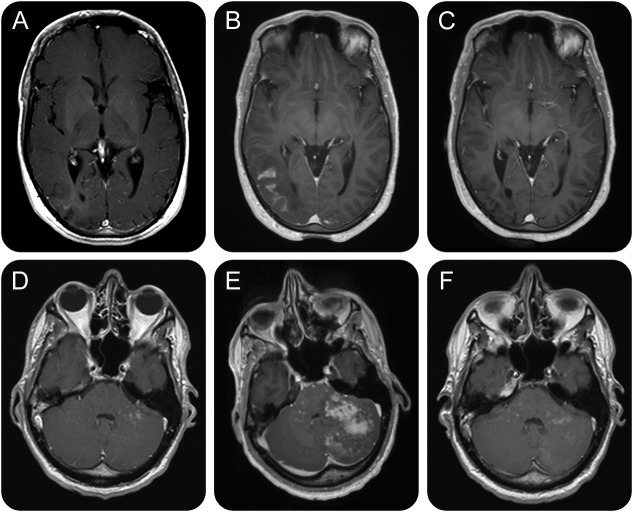
Figure. Serial MRIs of the brain.
Serial axial postcontrast T1-weighted magnetic resonance images of the brain of case 1 (A–C) and case 2 (D–F). (A) In February 2015, a T1-weighted image showed right subcortical occipital lesion (hyperintense in T2-weighted images) with subtle contrast enhancement suggestive of progressive multifocal leukoencephalopathy (PML) in a clinically asymptomatic patient. (B) Four months later, MRI showed progression of the contrast-enhancing occipital lesion, and maraviroc treatment was initiated. (C) Six months after start of maraviroc, MRI shows regression of PML–immune reconstitution inflammatory syndrome (IRIS) with no detectable contrast enhancement. (D) A routine MRI in February 2015 showed disseminated contrast-enhancing lesions in the cerebellum suggestive of PML in a clinically asymptomatic patient. (E) In May 2015, a T1-weighted image showed extensive IRIS after discontinuation of maraviroc. (F) Eleven months after the initial diagnosis of PML and after 8 months of maraviroc treatment, multiple disseminated contrast-enhancing lesions in the cerebellum are still detectable.

