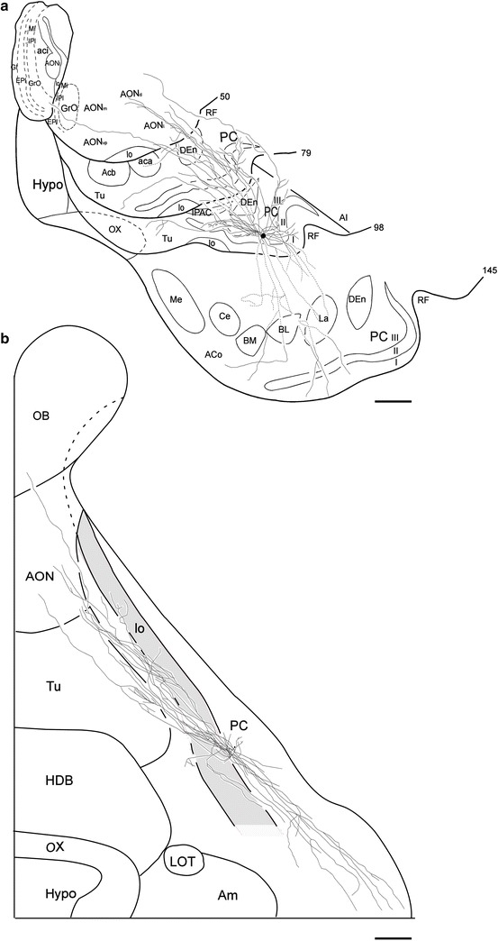
Fig. 5.

Coronal (a) and surface (b) views of reconstructed axon collaterals of a superficial pyramidal cell (SP-2 cell) located in the left hemisphere APC. Compared with the collaterals of the SP-1 cell (Fig. 4), the collaterals of this cell are very similar. a The axon collaterals are widely distributed within the PC and other olfactory areas and a long axon collateral projects to the olfactory bulb. b The small dot indicates the position of the soma. The collaterals extend in both rostral and caudal directions. The thick solid lines indicate the border between the areas of the cortical surface. Scale bar = 1 mm
