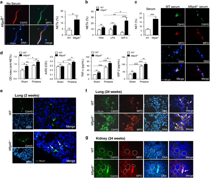
Figure 5.
MFG-E8-deficient neutrophils were primed to NETs release. (a) BMDNs from the Mfge8−/− or WT mice spontaneously release NETs without serum cultivation. Representative fluorescence microscopic images showing NETs. DNA was stained blue, citrullinated histone H3 was stained green and MPO was stained red. Three colors were merged by software Image J (National Institutes of Health, Bethesda, MD, USA), original magnification × 400. NET%=positive enlarged nuclei/total neutrophils per field. The average of NET release ratio was evaluated >500 cells in more than five fields per sample were counted. (b) PMA, LPS and MIP-2 treatment induced NETs formation from the Mfge8−/− and WT BMDNs. (c) The serum from pristane-treated mice stimulated NETs release. WT BMDNs were incubated with 2% serum isolated from the Mfge8−/− or WT, which were exposed to pristane for 24 weeks mice. Representative fluorescence microscopic images showing NETs release. (d) The serum levels of anti-NET antibodies, CICs, TNF-α and MIP-2 in Mfge8−/− and WT mice after pristane exposure for 24 weeks were measured by ELISA. (e) In vivo evidence for NETs formation in the lung tissues of Mfge8−/− mice after pristane exposure for 2 weeks. NETs were identified by close localization of DNA (blue) with citrullinated histone H3 (green), white arrows indicated NETs. (f) NETs were visualized in the lung tissues of Mfge8−/− or WT mice after pristane exposure for 24 weeks. (g) NETs were located around glomeruli in kidney tissues of Mfge8−/− mice after treatment with pristane for 24 weeks. Enlarged nuclei were founded to be citrullinated histone H3 and MPO positive, white arrows indicated NETs. For all experiments, data are shown as the means±S.E.M., *P<0.05, **P<0.01,***P<0.001; ns, not significant

