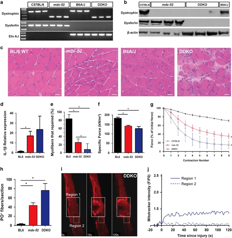
Figure 4.
Dysferlin is essential for sarcolemmal repair of dystrophin-deficient myofibers. C57BL/6 DDKO were generated by crossing mdx-52 and B6A/J mice and confirmed by (a) genomic DNA PCR and (b) WB analysis. (c) Images showing histological features of TA muscles from 28-day-old mice. Note the significantly enhanced muscle pathology in DDKO muscles. Scale bar=20 μm. (d) Quantification of the relative expression of interleukin IL-1β in 28-day-old quadriceps muscles from BL/6 WT, mdx-52, and DDKO. (e) Myofiber repair following laser injury was quantified in 29 DDKO myofibers (from 3 mice). Only 8±4% of the DDKO myofibers were able to repair compared with 21±8% of mdx-52. (f) Contractile force generation by the EDL muscle from 28-day-old BL/6 WT, mdx-52, and DDKO mice. (g) Loss in muscle contractile force following mechanical injury by 10% LC was monitored for EDL muscle from 28-day-old BL/6 WT, mdx-52, and DDKO mice. Note the greatest loss in contractile force in DDKO mice. (h) Quantification of PO-labeled myofibers following nine LC injuries was significantly greater in DDKO than mdx-52 mice. (i) Time-lapse images of mitochondrial dynamics during focal laser injury of DDKO sarcolemma within the region marked by the box with solid line (Region 1). Region 2 (box with dotted line) marks a region away from the injury site. (j) Quantification of mitochondrial accumulation indicated by MitoTracker intensity. Note that mitochondria fail to accumulate at the site of injury (Region 1). *P≤0.05 by one-way analysis of variance (with Bonferroni correction posttest)

