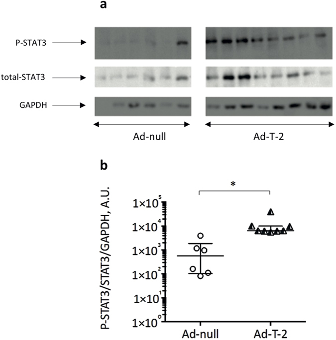
Figure 5. Determination of lung STAT3 expression and activation following sequential i.n Ad-null/Ad- T-2 and i.p Pb-ANKA administrations.

The lung samples (each one representing a different animal) obtained for Fig. 4 analysis were also assessed for expression of total STAT3, phosphorylated (P)-STAT3 and GAPDH. Lung extracts were analysed by SDS-PAGE, followed by Western Blot analysis. The PVDF membrane was incubated with rabbit anti-STAT-3 or anti-P-STAT3 antibodies (diluted 1:2,000) whereas anti-GAPDH antibody (to check for equal loading) was diluted 1:10,000. After incubation with secondary antibodies, the membrane was developed as described in Methods. The gel presented is a cropped version of the original gels presented in Fig. 3 Supplementary. (B) The ratios of P-STAT3/STAT3/GAPDH were determined (A.U = arbitrary units) and used as an indication of STAT3 activation. *Indicates a significant difference between groups (Student t test, p = 0.0007).
