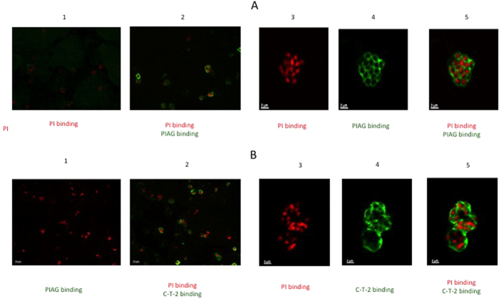
Figure 7. Illustration of C-T-2 and PIAG colocalization with merozoites.
(A) Co-localization of parasite immune IgGs (PIAG) with merozoites. Merozoite bags were prepared as indicated in Methods and incubated or not with PIAG. Nuclei were identified with PI binding (red fluorescence, low and high magnification, panels 1–2 and 3–5, respectively). Binding of PIAG with merozoites (green fluorescence) is shown both at low (panel 2) and high (panels 4–5) magnification. (B) localization of C-T-2 within merozoites. Merozoite bags were incubated with 10 μM of recombinant human C-T-2 (aa 38–95 from the full length T-2 molecule), as indicated in Materials and Methods. After permeabilization with acetone/methanol (50:50 dilution), rabbit anti-T-2 IgG (1:50 diution) and goat anti-rabbit IgG (1:500 dilution) coupled with Alexa 488 (green fluorescence) were added sequentially and incubated for 1 hr. As above, nuclei were identified with PI binding (red fluorescence, low and high magnification, panels 1–2 and 3–5, respectively). Binding of C-T-2 on merozoites (green fluorescence) is shown both at low (panel 2) and high (panels 4–5) magnification.

