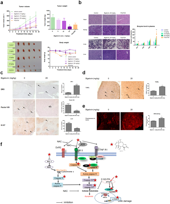
Figure 6. Bigelovin suppressed tumor growth of HCT 116-derived tumor xenografts in nude mice model.
HCT 116 cells were subcutaneously injected into the back of nude mice. Mice were treated with vehicle control, bigelovin or FOLFOX. Mean ± SEM; *p < 0.05, **p < 0.01, ***p < 0.001 vs. control at the same time point. (a) Tumor growth curve and body weight were calculated twice a week, tumor weight was measured at the end of the experiment. Representative data for 4–5 tumors in each group. (b) Organs were examined by H&E staining and plasma enzyme activities of ALT, AST, LDH and CK were calculated at the end of the experiment. (c) Tumor tissues were examined by IHC staining with antibodies against DR5, factor VIII and Ki67. (d) TUNEL analysis of bigelovin effects on tumor tissues in control and 20 mg/kg bigelovin treatment group. Representative images of 4–5 samples from each group in paraffin-embedded tissue sections. (e) Detection of superoxide levels by DHE staining in cryosections from tumors. The mean fluorescent intensity was obtained from 2–3 random visual fields of each tumor and quantified by ImageJ software. (f) The putative working model of bigelovin against colorectal cancer: bigelovin induces ROS and increases DR5 expression, then activate downstream caspase and cause G2/M and DNA damage through regulating p21, p-Rb and p-H2AX expression.

