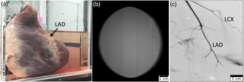
Figure 3. MuCLS angiography image.
(a) Photograph of the sample in waterbath. (b) Empty image of full MuCLS beam. (c) Quasi-mono-energetic angiography image of a porcine heart acquired at the MuCLS, with iodine-based contrast agent injected into the left coronary artery. Visible are the left anterior descending artery (LAD) and the left circumflex artery (LCX).

