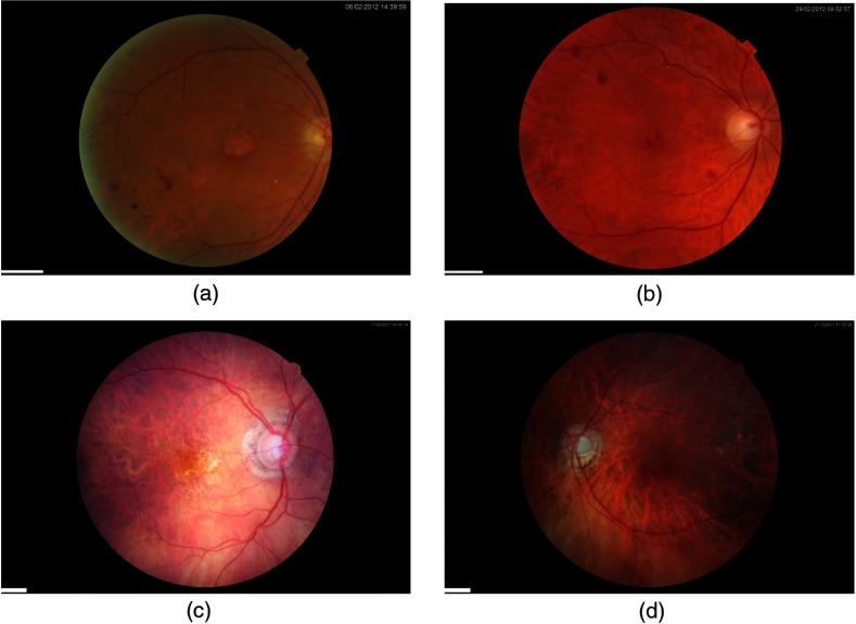
Fig. 3.
Example fundus images from the DR HAGIS database. The DR HAGIS fundus image database consists of four comorbidity subgroups. Fundus images of each of these four subgroups are shown here. (a) Diabetic retinopathy, (b) hypertension, (c) AMD, and (d) glaucoma. The scale bars (short white bar in the bottom left corner of each panel) correspond to 300 pixels.

