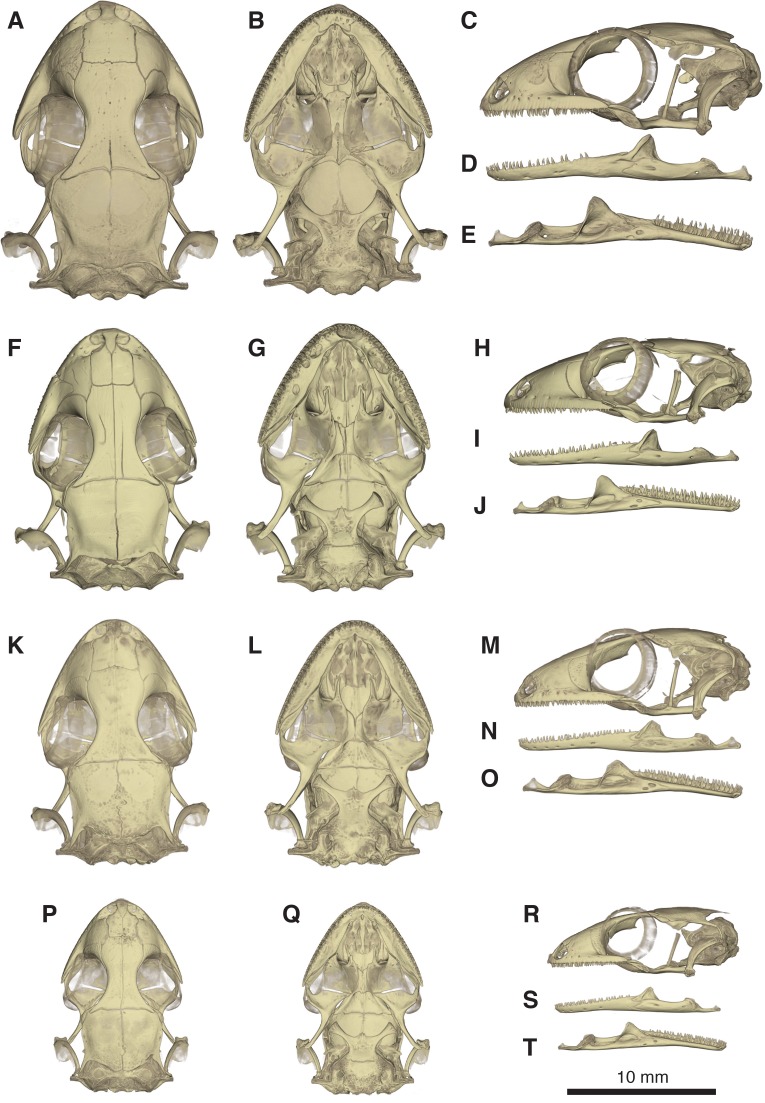
Figure 10. Comparative micro-CT images of the skull (cranium and jaw) of other Geckolepis species.
Shown in dorsal (A, F, K, P), ventral (B, G, L, Q), lateral (C, H, M, R), labial (D, I, N, S), and lingual (E, J, O, T) view. Depicting Geckolepis OTU AB (A–E, ZSM 1520/2008), holotype of G. maculata (F–J, ZMB 9655), and G. humbloti (K–O, ZSM 80/2010; P–T, ZSM 81/2006). From volume-rendering of micro-CT scans. Rotational videos of these skulls are provided in Videos S2–S5. For labels, see Fig. 6.

