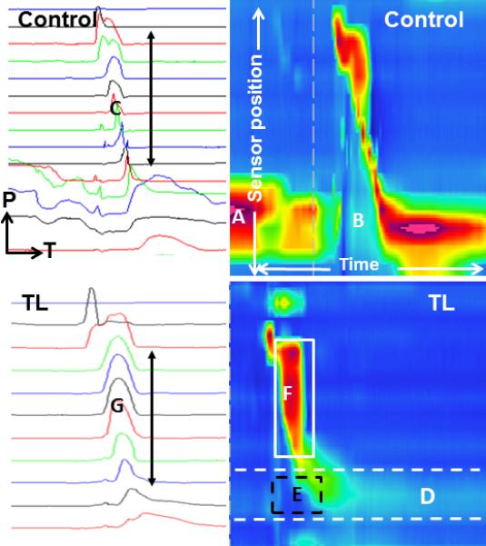
Figure 5.

Comparison of two-dimensional and spatiotemporal plots obtained from a control subject (above) and total laryngectomy subject (below). Two-dimensional plots are on the left, with each line representing one pressure tracing from one sensor; pressure (P) is on the y-axis, time (T) is on the x-axis, and more caudal sensors are at the bottom of the image. Spatiotemporal plots are on the right, with sensor position on the y-axis, time on the x-axis, and pressure represented by color. Each image represents 5 seconds of data collection. The vertical dashed line in the upper spatiotemporal plot is an artifact marking a time window from the data collection program. The control subject demonstrates a high resting upper esophageal sphincter (UES) pressure (A), low nadir pressure during bolus passage (B), and variation in shape of the pressure curves generated along the pharynx (C). The total laryngectomy subject demonstrates low resting UES tone (D, dashed white lines), maintenance of positive UES pressure during bolus passage (E, dashed black box), and a common cavity pressure (F, solid white box) which manifests as uniform pressure peaks along the neopharynx (G).
