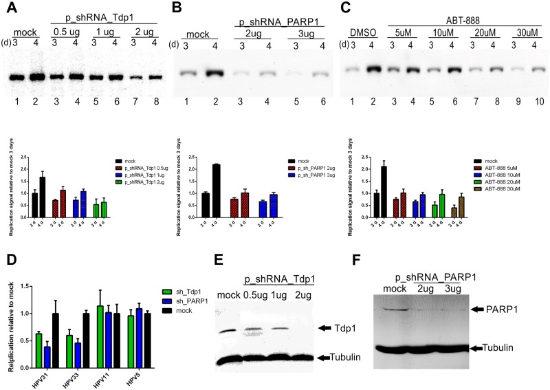
Fig 5. Tdp1 and PARP1 are essential cellular proteins for the initial amplification of the high-risk HPV genome.
U2OS cells were transfected with HPV18 wt minicircle and an sh_Tdp1 plasmid (A) or sh_PARP1 plasmid (B). Genomic DNA was extracted 3 and 4 days after the transfection, linearized and digested with DpnI. The HPV18 replication signal was detected with Southern blot analyses and quantified with a phosphoimager. C: U2OS cells were transfected with an HPV18 wt minicircle and grown for 3 and 4 days in the presence of different concentrations of the PARP1 inhibitor ABT-888. The HPV replication signal was detected with Southern blot analyses and quantified with a phosphoimager. D: U2OS cells were transfected with an HPV31, 33, 11 or 5 wt minicircle and the sh_Tdp1 or sh_PARP1 plasmid (2μg). Genomic DNA was extracted 3 days post-transfection, linearized and digested with DpnI The HPV replication signals were detected with Southern blot analyses and quantified with a phosphoimager. E and F: Western blot analyses showing the downregulation of the Tdp1 and PARP1 proteins by shRNA at the 3-day timepoint. Empty shRNA vector was used as a mock control for Tdp1 and PARP1 downregulation by sh_RNA plasmids. G: Error bars represent standard deviations from two to three independent experiments.

