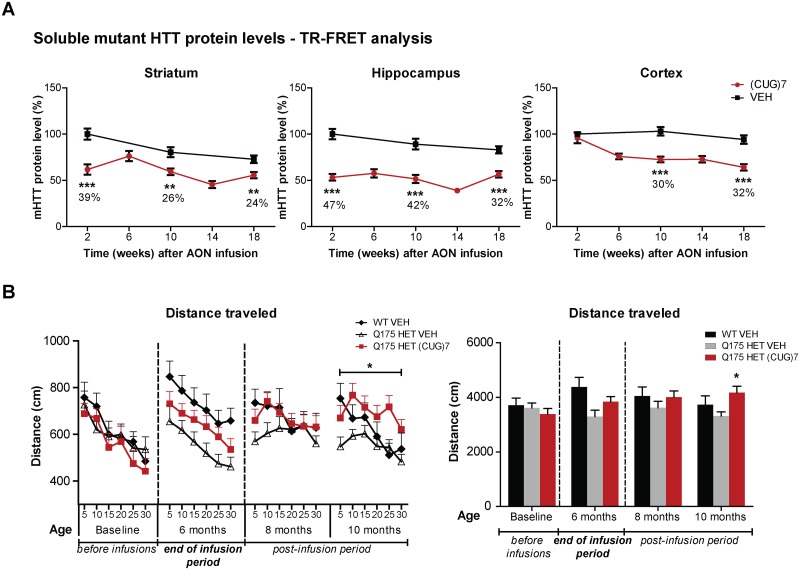
Fig 6. Effects of (CUG)7 treatment in the Q175 HD mouse model.
(A) Levels of soluble mHTT protein in striatum, hippocampus and cortex, as determined by TR-FRET-based immunoassay [22]. Each point is the average of 3 technical replicates. Soluble mHTT protein levels are expressed relative to levels after VEH-treatment at the 2 week post infusion time point (set to 100%). Data are presented as mean ± SEM. Significance was assessed using a 2-tailed t-test comparing (CUG)7-treated mice to VEH controls per time point (**p<0.01, ***p<0.001). (B) Distance traveled in the open field at baseline and different time points post infusion. Mice were placed in the center of the chamber and their behavior was recorded for 30 min in 5-minute bins (individual bins shown in left graph, cumulative distance in right graph). A significant increase in distance traveled was observed in 10-month old (CUG)7-treated Q175 mice, which corresponds to 4 months post last infusion (around the 18 week post infusion time point). Data are presented as mean ± SEM. Significance was assessed using a 2-tailed t-test comparing (CUG)7- treated Q175 mice to VEH controls per time point (*p<0.05).

