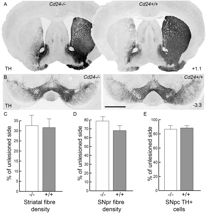
Fig 4. The absence of Cd24 has no effect on dopaminergic neuronal survival or fibre innervations in the striatal 6-OHDA lesioned mouse model of Parkinson's disease.
(A) Tyrosine hydroxylase (TH) immunohistochemistry on coronal sections of brain from a 6-OHDA lesioned Cd24-/- mouse (left column) and a Cd24+/+ littermate (right column) at 12 days post-surgery, at the level of the striatum. (B) Representation sections of midbrain (from the respective brains provided in panel A) illustrating the loss of TH+ cells on the 6-OHDA lesioned side of the brain. (C) Optical density analysis of the TH+ fibres in the striatum indicated no difference between the genotypes. (D) Optical density analysis of the TH+ fibres in the SNpr also showed no significant difference between the genotypes. (E) Stereological estimations of the number of SNpc TH+ cells found no difference between the Cd24-/- and Cd24+/+ mice. Co-ordinates in the right-hand corners of panels A indicate location of the coronal plane relative to bregma. The scale bar in panel A represents 1cm and 500μm in panels B.

