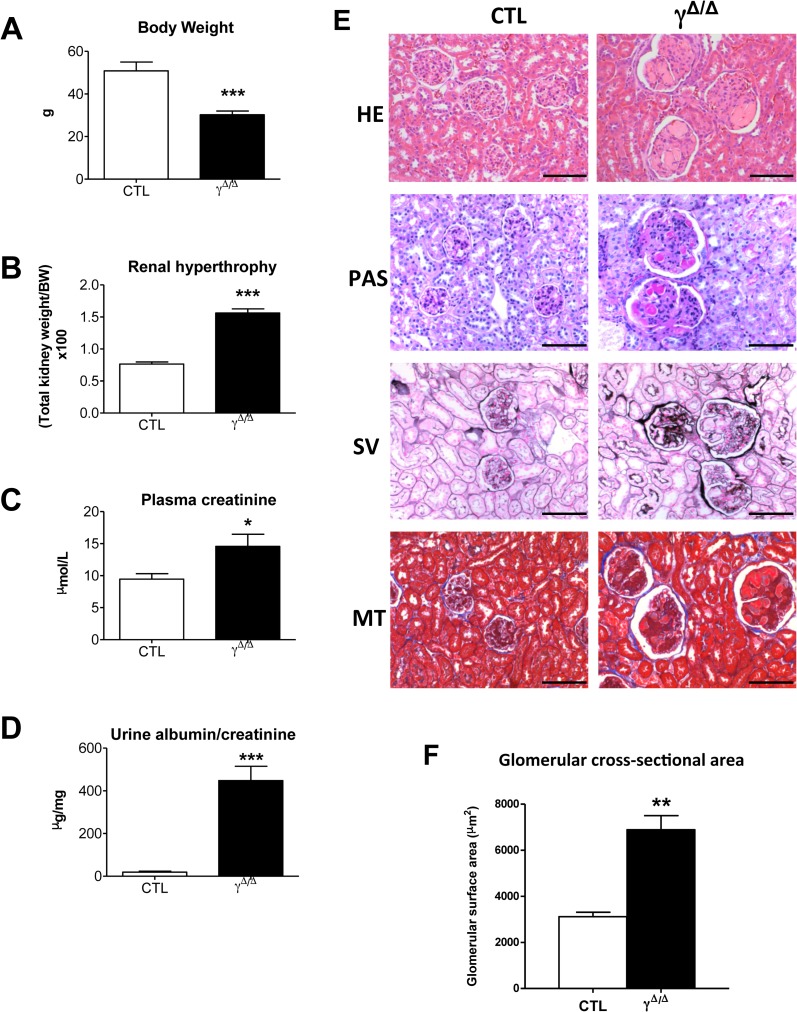
Fig 2. Chronic functional and histological damages in ageing PpargΔ/Δ kidney.
Indicated parameters and histological studies were performed in 52 weeks old control (CTL) and PpargΔ/Δ (γΔ/Δ) mice. (A) Body weight (6 controls and 9 PpargΔ/Δ P = 0.0002), (B) renal hypertrophy as evaluated by the ratio total kidney weight over total body weight (X100) (6 controls and 9 PpargΔ/Δ P<0.0001), (C) plasma creatinine (6 controls and 7 PpargΔ/Δ P = 0.0384), (D) albuminuria (7 controls and 9 PpargΔ/Δ P<0.0001) (E) Representative images of paraffin kidney sections stained with haematoxylin and eosin (HE), Periodic Acid Shiff (PAS), Silver methenamine (SV), and Masson’s trichrome (MT). Scale bar represents 100μm. (F) glomerular cross-sectional area in 52 weeks female mice (4 controls and 5 PpargΔ/Δ P = 0.0011). Data in the graphs corresponds to the mean ± SEM. *P<0.05, **P<0.01, ***P<0.001 PpargΔ/Δ vs. age matched control littermates.

