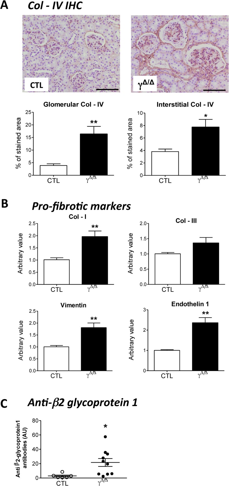
Fig 3. Immunohistochemistry and gene expression of fibrosis markers in kidney of ageing PpargΔ/Δ mice, and plasmatic levels of anti-β2-glycoprotein1 antibodies.
52 weeks old control (CTL) and PpargΔ/Δ (γΔ/Δ) animals were analyzed for the following parameters: (A) Top panels: Kidney Immunohistochemistry for collagen IV (Col-IV). Positive staining is in brown. Scale bar represents 100μm. Bottom panels: quantification of Col-IV deposition at glomerular levels (4 controls and 4 PpargΔ/Δ P = 0.0067) and tubulointerstitial levels (4 controls and 4 PpargΔ/Δ P = 0.0238), (B) Evaluation by RT-qPCR of the following gene expression in the kidney of CTL (N = 5) and PpargΔ/Δ (N = 7) mice: collagen I (Col-I, P = 0.0081), collagen III (Col-III, P = 0.1441, not significant), vimentin (P = 0.0083) and Endothelin 1 (P = 0.0015). Results are reported as fold change with respect to control levels, which were arbitrarily set to 1. (C). Plasma levels of antibodies against the β2-glycoprotein1 (6 controls and 10 PpargΔ/Δ P = 0.0233). Data in the graphs corresponds to the mean ± SEM. *P<0.05, **P<0.01 PpargΔ/Δ vs. age matched control littermates.

