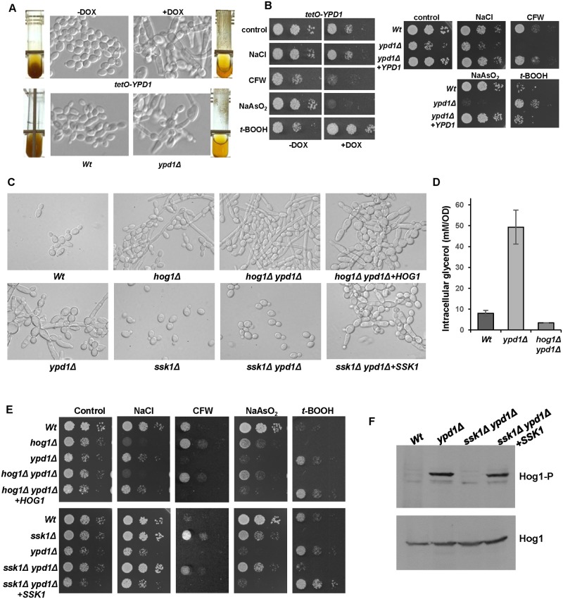
Fig 3. Phenotypes associated with loss of Ypd1 are dependent on Hog1 and Ssk1.
(A) Repression or deletion of YPD1 triggers flocculation and a swollen pseudohyphal filamentous phenotype. Micrographs of Wt, ypd1Δ, and tetO-YPD1 cells plus or minus doxycycline (DOX) grown overnight in rich media. Images of culture tubes demonstrate the rapid sedimentation rate of cells lacking YPD1. (B) Repression or deletion of YPD1 results in pleiotropic stress phenotypes. 104 cells, and 10-fold dilutions thereof, of exponentially growing tetO-YPD1 cells, or wild-type (Wt), ypd1Δ and ypd1Δ+YPD1 cells, were spotted onto rich media plates (plus or minus DOX for tetO-YPD1 cells) containing NaCl (1.0 M), calcofluor white (CFW, 30 μg/ml), NaAsO2 (1.5 mM) and t-BOOH (2 mM), and incubated at 30°C for 24h. (C) The morphological defects exhibited by ypd1Δ cells are dependent on Hog1 and Ssk1. Micrographs of wild-type (Wt), ypd1Δ, hog1Δ (JC50), ssk1Δ (JC1552), hog1Δ ypd1Δ (JC1475) hog1Δypd1Δ+HOG1 (JC1478), ssk1Δ ypd1Δ (JC1683), and ssk1Δ ypd1Δ+SSK1 (JC1704) cells. (D) The high glycerol levels in ypd1Δ cells are dependent on Hog1. The mean ± SD is shown for 3 biological replicates. (E) The stress phenotypes exhibited by ypd1Δ cells are dependent on Hog1 and Ssk1. Exponentially growing strains were spotted onto rich media plates containing the additives detailed in B above, and incubated at 30°C for 24h. (F) The sustained Hog1 activation in ypd1Δ cells is dependent on Ssk1. Western blots depicting basal levels of Hog1 phosphorylation in the indicated strains. Blots were probed for phosphorylated Hog1 (Hog1-P), stripped and reprobed for total Hog1 (Hog1).

