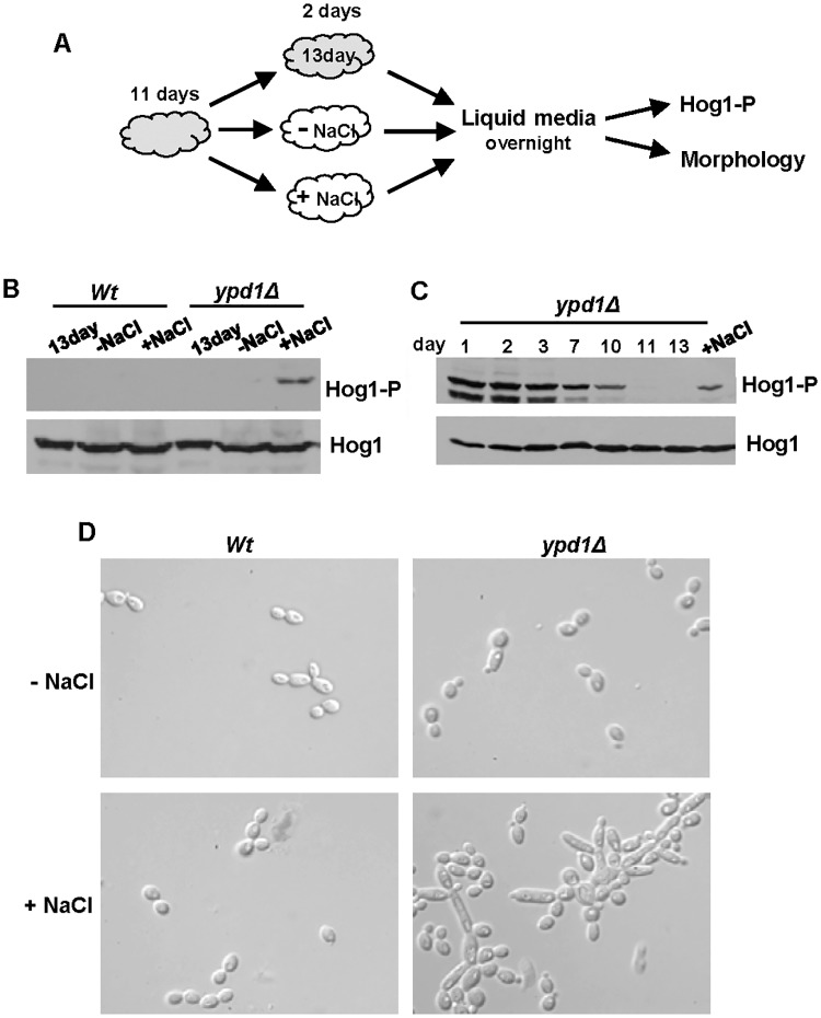
Fig 6. The reduction of Hog1 phosphorylation following loss of Ypd1 function can be reversed by stress exposure.
(A) Experimental overview. Freshly isolated cells were incubated on rich solid media for 11 days (depicted in grey) and then either re-streaked onto fresh rich media with (+NaCl) or without (-NaCl) 0.3M NaCl (depicted in white) and incubated for a further 2 days, or maintained on the original plate for a further 2 days (13 day). Cells were then cultured overnight in liquid rich media lacking NaCl, and Hog1 phosphorylation and cellular morphology examined. (B) Western blot analysis of both phosphorylated and total Hog1 levels in Wt (JC21) and ypd1Δ (JC2001) cells treated as described in A. (C) Western blot analysis of whole cell extracts isolated from exponentially-growing ypd1Δ cells taken either from rich media plates after the number of days indicated, or after being re-streaked on rich media with NaCl (+NaCl) as described in A. Duplicate blots were probed for phosphorylated Hog1 (Hog1-P) or total Hog1 (Hog1) levels. (D) Micrographs of exponentially-growing Wt and ypd1Δ strains treated as described in A.

