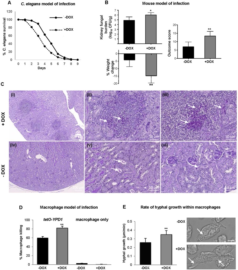
Fig 7. Repression of YPD1 expression during infection potentiates C. albicans virulence.
(A) C. elegans model of infection. Nematodes were infected with the conditional tetO-YPD1 strain (JC1586) and transferred to liquid medium either with (+DOX) or without (-DOX) doxycycline. Doxycycline treatment consistently increased the rate of killing of the nematodes infected with the tetO-YPD1 strain (P<0.001). These data are from a single experiment representative of three independent biological replicates. (B) Mouse model of infection. Kidney fungal burden measurements, percentage weight loss, and outcome score measurements of mice infected with tetO-YPD1 cells and administered doxycycline (+DOX) or not (-DOX). Comparison of +DOX and -DOX treated groups by Kruskal-Wallis statistical analysis demonstrates significant differences for all three parameters with doxycycline treated mice giving a significantly greater outcome score (*P<0.05, ** P<0.01). (C) Increased virulence of cells with repressed YPD1 expression is associated with increased numbers of fungi and inflammation in the infected kidneys. Representative images from kidneys of mice infected with tetO-YPD1 cells and treated with (+DOX; i-iii) or without (-DOX; iv-vi) doxycycline. Sections (5 μm) were Periodic Acid-Schiff stained and post-stained with Hematoxylin. Low magnification images in (i) and (iv) (scale bar = 200 μm) show the difference in lesion number and extent of inflammatory response. Higher magnification (scale bar = 50μm) shows the presence of clusters of filamentous fungal cells (white arrows) in the lesions of doxycycline-treated mouse kidneys (ii & iii) and isolated single pseudohyphal fungal cells (white arrows) in placebo-treated mouse kidneys (v & vi). (D) Macrophage model of infection. C. albicans mediated killing of macrophages was determined by detecting the number of ruptured macrophages following co-culture with or without tetO-YPD1 cells in the presence (+DOX) or absence (-DOX) of doxycycline. ANOVA was used to determine statistical significance (** P ≤ 0.01). (E) Assessment of hyphal growth following phagocytosis by tetO-YPD1 cells in the presence (+DOX) or absence (-DOX) of doxycycline. The left panel shows that +DOX treatment resulted in a faster rate of hyphal growth. ANOVA was used to determine statistical significance (** P ≤ 0.01). The right panel shows images taken 61 mins post engulfment of yeast cells. The scale bar is 9 μm, and the white arrows indicate hyphal cells within the macrophage.

