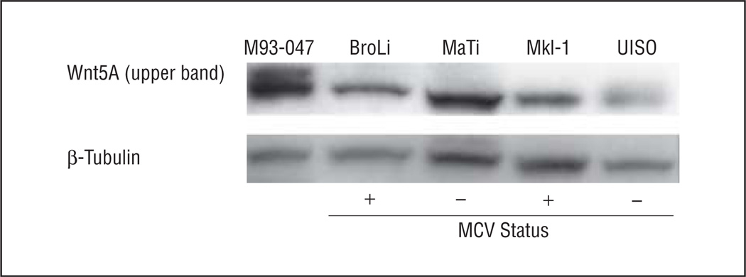
Figure 1.
Western blot analysis of Wnt5A expression in Merkel cell cancer cell lines. The indicated cell lines were lysed in radioimmunoprecipitation assay buffer and subjected to Western blot analysis. Wnt5A (upper band) is present in the melanoma cell line M93-047, used as the positive control, and registers faintly positive in the UISO cell line. β-Tubulin was used as a loading control. MCV indicates Merkel cell polyomavirus. Plus signs and minus signs indicate positive and negative MCV status, respectively.

