. 2017 Jan 12;6:e17681. doi: 10.7554/eLife.17681

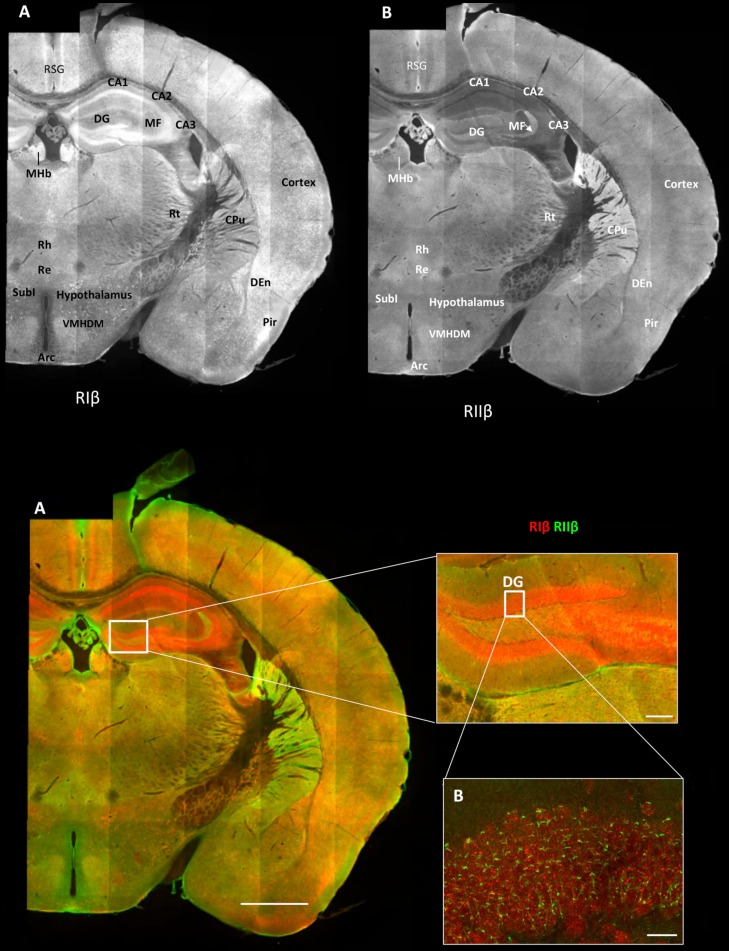
Figure 1. Overview of the regional distribution of RIβ and RIIβ across brain regions.

Wide-field views of a high-resolution large-scale mosaic image for a full coronal tissue slice are shown for RIβ (red in colored image) and RIIβ (green in colored image). The tissue section was labeled with anti-RIβ- and anti-RIIβ-specific antibodies. The mosaic image was made up of 105 tiles. Each tile is a maximum intensity projection of a stack of 15 Z-sections that were stitched together to reconstruct this single, high-resolution 2D image. Colored image: (C) Scale bar inside the full mosaic image: 1 mm. Scale bar inside the small box: 100 µm. (D) Intermediate resolution sample. (E Full-resolution sample of the mosaic allows examination at higher magnification. White boxes represent the areas from which the image was captured. DG, Dentate Gyrus. Scale bar: 10 µM. Abbreviations: Arc, arculate nucleus; CPu, Caudate putamen; DEn, dorsal endopiriform nucleus; MF, mossy fibers; MHb, medial habenular nuclear; Pir, piriform cortex; Re, reuniens thalamic nu; Rh, rhomboid thalamic nucleus; RSG, retrosplenial granular cortex; RSV, retrosplenial granular; Rt, reticular thalamic nucleus; SubI, subincertal nucleus.

DOI: http://dx.doi.org/10.7554/eLife.17681.002

Figure 1.

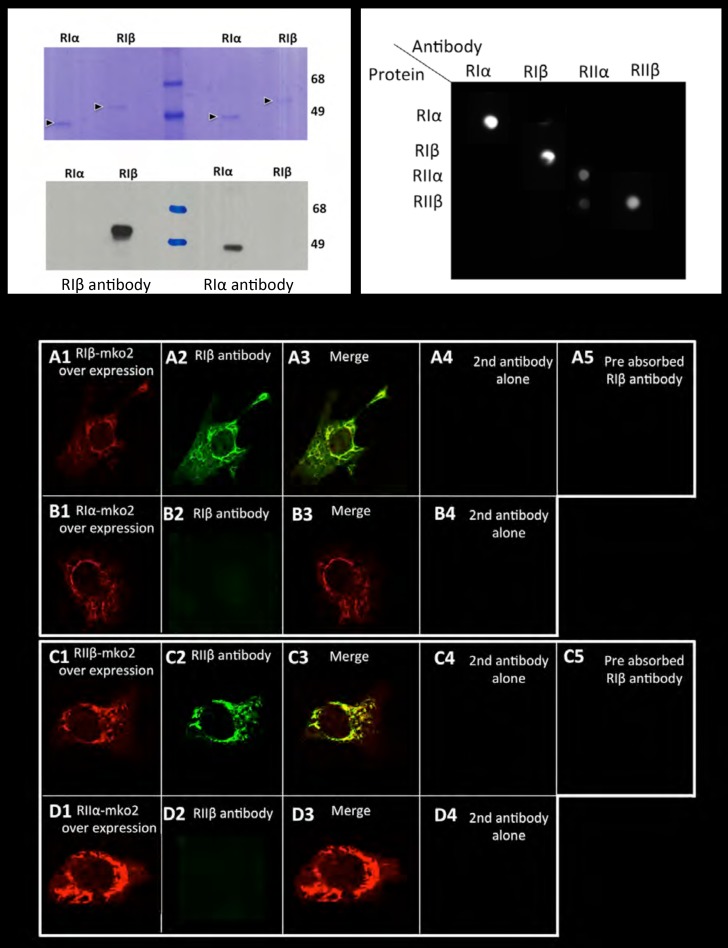
Figure 1—figure supplement 1. Antibody specificity.

Figure 1—figure supplement 1.

Left panel: Commassie blue stained gel or western blot of purified RIα/RIβ proteins detected by RIα/RIβ antibodies. Right panel: Dot blot analysis. Each row of the nitrocellulose membrane was spotted with all four purified PKA regulatory subunits as indicated, followed by specific antibody detection. Lower panel: 10 T1/2 mouse cell cultures were transfected with (A) RIβ-MKO2 or (B) RIα-MKO2 and were immunostained with RIβ antibody (A2, B2). Merge images are shown (A3, B3). No unspecific staining was detected in controls for secondary antibodies alone (A4D4). Pre-absorption of RIβ or RIIβ antibodies with purified RIβ or RIIβ proteins, respectively, resulted in blocking the antibody signal (A5C5). A similar experiment was carried out with overexpressing RIIβ-MKO2 (C1) or RIIα-MKO2 (D1). Cells were immunostained with RIIβ antibody (C2, D2). Merge images are shown in (C3D3). No unspecific staining was detected in controls for secondary antibodies alone (C4, D4).
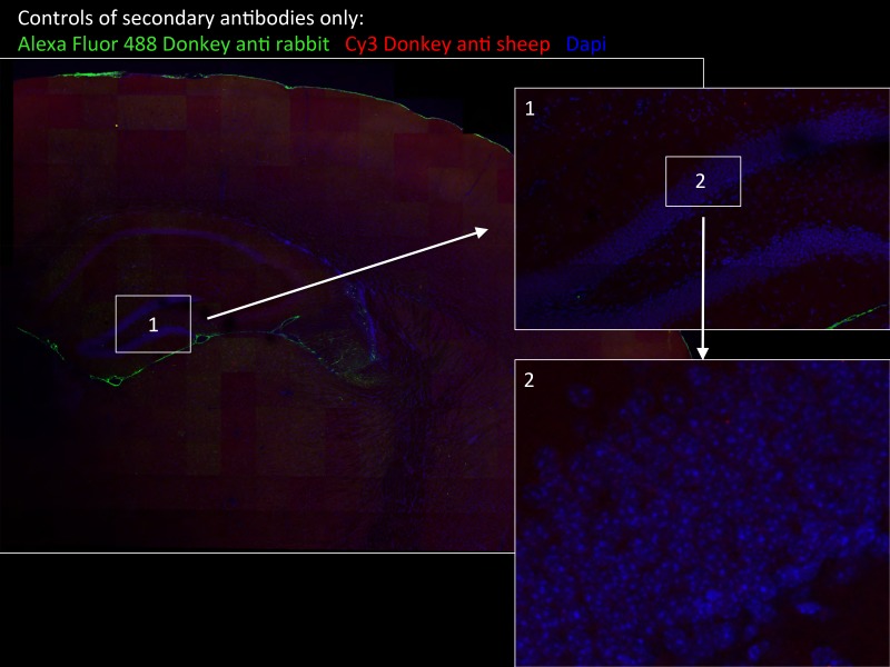
Figure 1—figure supplement 2. Specificity of secondary antibodies.

Figure 1—figure supplement 2.

Wide-field views of a high-resolution large-scale mosaic image for a full coronal tissue slice are shown for secondary antibodies alone: Alexa Fluor 488 donkey anti-rabbit (green) and CY3 Donkey anti-sheep (red) including dapi staining (blue). (2) Full-resolution sample of the mosaic allows examination at antibody controls at higher magnification. No staining was detected. White boxes represent the areas from which (1) and (2) was captured. DG, dentate gyrus.