. 2017 Jan 12;6:e17681. doi: 10.7554/eLife.17681

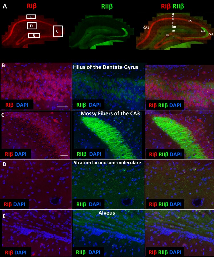
Figure 3. RIIβ is the predominant isoform in cell axons across various hippocampal subfields.

(A) A full representative mosaic of a coronal section through the dorsal hippocampus at reduced resolution. An overview of RIβ (red), RIIβ (green) and RIβ/RIIβ immunostaining at various subfields. The mosaic image is made up of 1,413 tiles from ten Z sections obtained using a 60x objective lens. The white box represents the area from which the (BE) subfield full-resolution images were captured. (B) Hilus of dentate gyrus. (C) Mossy fiber of the CA3. (D) Stratum lacunosum-moleculare. (E) Alveus. Abbreviations: CA1, stratum oriens; g, granule cell layer (stratum granulosum); h, hilus proper; lm, stratum lacunosum-moleculare; m, dentate molecular layer (stratum moleculare); p, stratum pyramidale; r, stratum radiatum. Scale bar: 25 µm.

DOI: http://dx.doi.org/10.7554/eLife.17681.006

Figure 3.

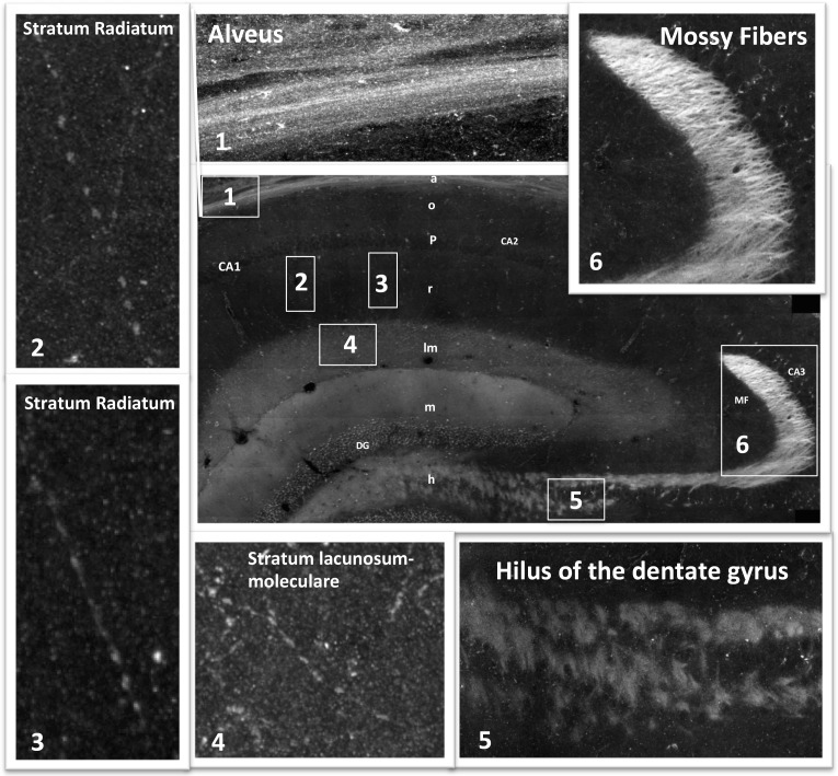
Figure 3—figure supplement 1. Mosaic images of the hippocampus allow tracing of RIIβ along an axonal pathway and visualization of axonal boutons in the full-resolution mosaic image.

Figure 3—figure supplement 1.

Middle image: Immunostaining of RIIβ in a full representative mosaic of coronal section through the dorsal hippocampus. Subfields of the hippocampus are marked as in the main Figure 3. Numbers in white boxes represents the area from which the full-resolution images (1–6) were captured. Images 5 and 6 show full-resolution images of the axonal pathway that emerges from the granule cells of the dentate gyrus and passes through the hilus to the mossy fibers at the CA3. Images 2, 3 and 4 show full-resolution images of RIIβ staining the axonal terminal boutons.