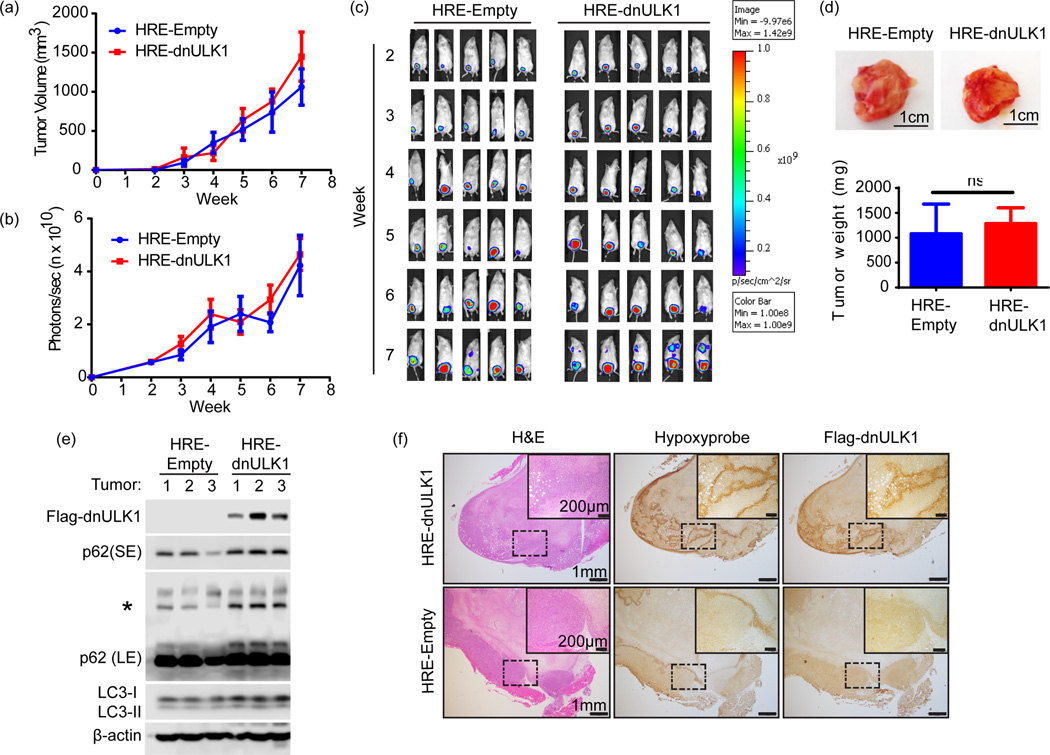
Figure 2. Loss of ULK1 function in the hypoxic tumor cells does not affect primary tumor growth.
a, Tumor volume of primary xenograft tumors over 7 weeks (mean ± s.d, n=15 mice per group). b, Quantification of primary tumor luciferase expression photon flux (photons/sec) in HRE-dnULK1 and HRE-Empty xenografts (mean ±SEM, n=15 mice per group). c, Representative images of luciferase expression from primary xenograft tumors over a 7 week time period. d, Representative images of tumors from HRE-dnULK1 and HRE-Empty orthotopic xenograft mice and quantification of final weight of excised primary tumors (mean ± s.d, n=15, ns=no significance, t-test). e, Immunoblot analysis of dnULK1 and p62 expression from 3 primary tumors from HRE-empty and HRE-dnULK1 xenografts (SE= short exposure, LE = Long Exposure, Asterisks (*)= p62 aggregate). f, Immunohistochemistry staining of primary tumors and lung serial sections from HRE-dnULK1 xenografts at week 7. Hypoxyprobe™ detects hypoxic areas within each tissue, which correlates to dnULK1 expression.

