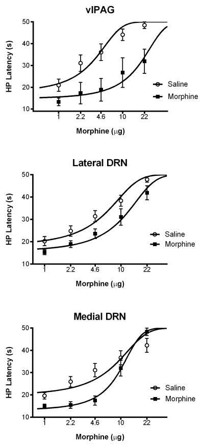
Figure 3.
Tolerance to morphine antinociception was greatest following microinjections into the ventrolateral PAG compared to the DRN. A significant rightward shift in the morphine dose response curve occurred in all three brain regions following repeated morphine microinjections. Changes in morphine potency were compared to saline pretreated rats and measured with the hot plate (HP) test. The magnitude of morphine tolerance was greatest with injections into the ventrolateral PAG (top) compared to the lateral (middle) or medial (bottom) DRN (see Table 1).

