
Figure 1. Immunohistochemical assessment of liver biopsies.
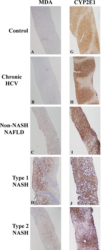
Representative images of lipid peroxidation levels [measured by malondialdehyde (MDA) staining] are shown in the left panels (A–F) and CYP2E1 staining is shown in the right panels (G–L) as follows: (A,G) Control; (B,H) Chronic HCV Infection; (C,I) non-NASH NAFLD; (D,J) Type 1 NASH; (E,K) Type 2 NASH; (F,L) Mixed NASH. Hepatic lipid peroxidation was significantly greater in pediatric NAFLD compared to controls and chronic HCV patients (compare panels A,B with panels C–F). Hepatic CYP2E1 protein content was not different in children with normal biopsies or chronic HCV infection versus those across the spectrum of NAFLD (compare panels G,H with panels I–L).