Figure 1.

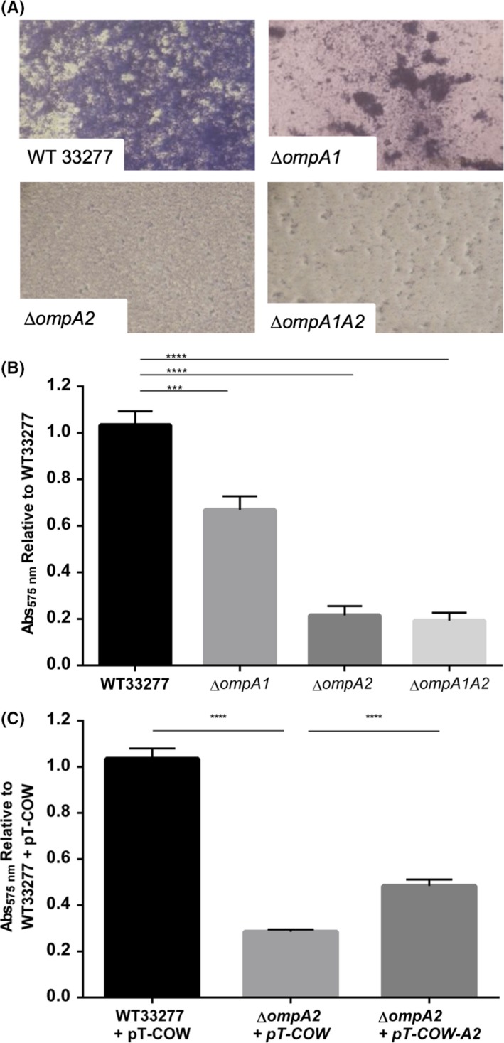
Biofilm formation in vitro. OD 600 nm 0.05 cultures were seeded and grown anaerobically for 72 hr, and biofilm stained with 1% Crystal Violet. Biofilms were imaged at 400× magnification (A), before Crystal Violet extracted and absorbance measured (OD 570) to quantify biofilm formation (B). The ∆ompA2 mutant was complemented and biofilm examined (C). Statistical significance was determined by students’ t‐test and designated as ***p < .001, **** p < .0001 (n = 3)
