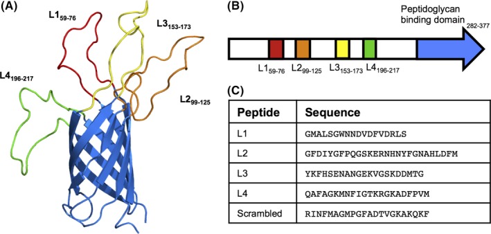
Figure 4.

In silico analysis of OmpA2 protein and extracellular loops. (A) Structure modeling of OmpA2, displaying transmembrane β‐barrel and predicted extracellular loops, L1‐L4. N‐terminal α‐helix and C‐terminal peptidoglycan domain have been removed for display purposes. (B) Schematic representation of the location of the extracellular loops (colour corresponding to β‐barrel image) and predicted peptidoglycan‐binding domain (pale green) in the ompA2 gene. Predicted extracellular loops sequences (C) were commercially ordered and Biotin‐tagged
