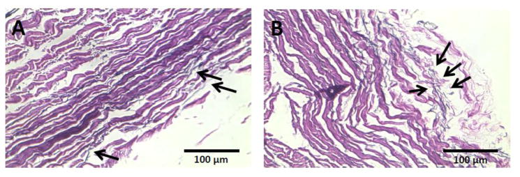
Fig. 11.
Verhoeff Van Gieson (VVG) staining of two cross sections extracted from Uncrimped (A) and crimped (B) bovine pericardial leaflets, respectively. Image A demonstrates intact elastin fibers (long black thin lines) in a cross section of an intact pericardial leaflet and image B shows fragmented elastin (short black thin lines) fibers in a stent-crimped leaflet. Elastin fibers in both cross sections were pointed by black arrows. Bars are 100 μm. The images are adapted from Sinha and Kheradvar (2015) 118

